Ultrasound, also called sonography or ultrasonography, uses high-frequency sound waves (above the audible range of the human ear) to produce medical images. Almost all parts of the body can be imaged with this versatile technique. In our practice, the most commonly examined areas are the female pelvic organs, fetus, breasts, thyroid gland, kidneys, gallbladder, and liver. We also perform sonograms of blood vessels (e.g. carotid arteries to look for plaques that may cause strokes, abdominal aorta to look for life-threatening aneurysms, or leg veins to look for clots that can fatally travel to the lungs), pancreas, spleen, appendix, testicles, and groin (for hernias).
Here are some sample sonogram images:
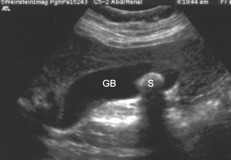
Stone (S) in the gallbladder (GB)
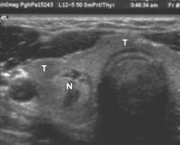
Benign nodule (N) in the thyroid gland (T)
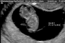
First trimester pregnancy
(head on top, rump on bottom)
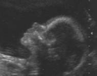
Second trimester pregnancy
(profile view of the face)
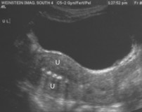
IUD (multiple white spots) in the middle of the uterus (U)