Mammography uses X-rays to produce pictures of the breast that can show signs of early breast cancer. "Screening" mammography is done when there are no breast problems and consists of two standard views of each breast. The American College of Radiology recommends screening mammography once a year beginning at age 40, unless there is a strong family history of premenopausal breast cancer, in which case screening may need to start earlier. Because cancers can develop between mammograms or not be visible on mammography, monthly breast self-examinations are also recommended.
"Diagnostic" mammography is performed when there are breast problems (e.g., newly discovered lump, pain, or bloody discharge from the nipple), a questionable abnormality on screening mammogram, or a mammographic abnormality that is being followed from a prior exam. Other indications for a diagnostic mammogram include prior breast cancer and breast implants. In addition to the two standard views, additional views with different positioning, different amounts of compression, or magnification may be necessary. If further evaluation is still needed after the additional views, breast ultrasound and/or biopsy may be warranted.
To date, mammography is the only test that has been proven to reduce breast cancer deaths. But it is also known that the accuracy of mammography decreases as the density of the breast tissue increases. Screening breast ultrasound, tomosynthesis (3D mammography), and breast MRI can be useful in women with dense breasts.
Digital Mammography
While traditional mammography uses film to capture an X-ray picture of the breast, digital mammography uses a digital sensor, similar to the sensor in a digital camera - only larger. Digital mammograms are viewed and interpreted on a high-definition computer monitor.
Researchers have found that digital mammography can be beneficial to women with dense breasts. More information about the Digital Mammographyic Imaging Screening Trial (DMIST) can be found on the National Cancer Institute site.
Tomosynthesis, also known as 3D mammography, uses a series of low-dose images to create three-dimensional pictures of the breasts. This allows the radiologist to view the breast tissue in layers without being hidden or obscured by dense tissue above or below each layer. The end result is (1) fewer additional mammographic images to determine whether a spot on the mammogram is due to a cancer or just overlapping shadows and (2) better detection of small cancers.
The options for women with dense breasts include:
1. Non-digital mammography with breast ultrasound (for women at high risk for breast cancer)
2. Digital mammography
3. Mammography (either type) with Breast-Specific Gamma Imaging (BSGI), a test that uses an intravenous tracing agent that accumulates more in cancers than in normal breast tissue
4. Breast MRI for women at high risk for breast cancer, which may be based on personal history or family history of breast cancer
Investigations are currently under way to determine which tests are best for various groups of individuals.
For additional information about mammography, please refer to RadiologyInfo.com.
Digital Breast Tomosynthesis
Because current regulations call for tomosynthesis to be performed in addition to a standard mammogram, there is extra radiation involved, though the total dose is still well below the maximum permissible amount for a standard mammogram. This is further improved by the reduced need for additional mammographic views. The hope is that tomosynthesis can completely replace 2D mammography in the not-too-distant future.
Breast Imaging at Weinstein Imaging Associates
Breast imaging and minimally invasive breast biopsy constitute a major focus of our practice. We strive to offer our patients high-quality examinations and accurate interpretations. We are pleased that statistics confirm that we're "doing the right thing," as we beat the national average in mammographic accuracy year after year. This is due in large part to the high level of specialized training attained by all our radiologists and the fact that we read large volumes of screening mammograms on a daily basis, which is important in maintaining mammographic interpretive skills.
Another component of a quality practice is quality equipment. All of our mammograms are performed digitally. We have offered digital breast tomosynthesis since August 2012. Our breast ultrasound exams are done on high-resolution, state-of-the-art units. We also provide Breast-Specific Gamma Imaging/ molecular breast imaging and 3D breast ultrasound.
Also of importance is the fact that we are involved with cutting-edge research, which is usually done by universities, but also by respected private groups such as ours. Our research projects have included breast ultrasound and MRI screening, as well as computer-aided detection for breast utlrasound with Medipattern Corporation.
Please contact usif you are interested in any of our breast imaging tests.
Mammography is useful for detecting calcifications (calcium deposits), which are sometimes a sign of cancer. On this mammogram, the white specks and lines are calcifications from widespread cancer.
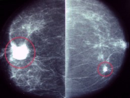
This woman had mammography after she felt a right breast lump. Mammogram revealed not only the cancer in the right breast (large circle in the left picture), but also an unsuspected cancer in the opposite breast (small circle in the right picture).
Copyright 2004-24 Weinstein Imaging Associates, Pittsburgh, PA
North Hills 412-630-2649 • Shadyside 412-441-1161 • South Hills 412-440-6999